Dubininaís (1982) monograph is based on a detailed anatomical and histological examination of the various species, which permits, for example, the distinction of a simple seminal receptacle and an accessory seminal receptacle. The key presented here is therefore based on Dubinina (1982), although it was strongly modified taking into account my own studies as well as the keys of Gibson (1994) and Bandoni and Brooks (1987).
Dubinina (1982) recognized four families, the Amphilinidae with two subfamilies, the Amphilininae (genus Amphilina) and Gephyrolininae (genus Gephyrolina), the Schizochoeridae (genera Schizochoerus and Nesolecithus), the Austramphilinidae (genus Austramphilina), and the Gigantolinidae (genus Gigantolina). Gibson (1994) distinguished two families, the Amphilinidae and Schizochoeridae, the former including the genus Amphilina, the latter the two subfamilies Schizochoerinae (genera Gephyrolina, Nesolecithus and Schizochoerus) and Austramphilinae (genera Austramphilina and Gigantolina). Differences between species of the families appear to be very slight. Furthermore, an egg stalk is found in genera included by Gibson in two families, i.e., in Amphilina and Gephyrolina (but in none of the others). Hence only a single family, Amphilinidae, is recognized here. Bandoni and Brooks (1987) synonymized Austramphilina with Gigantolina, and Nesolecithus and Gephyrolina with Schizochoerus. Further studies, particularly of the excretory system, egg and early cleavage, are necessary to justify these synonymizations, particularly in view of the fact that a polar egg stalk is present only in Amphilina and Gephyrolina, but not in any of the other genera, and that there are significant differences in the structure of the reproductive system of Austramphilina and Gigantolina. For a list of synonyms of the genus and species names used in the following see Synonyms.
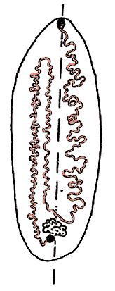
1.      Eggs with stalk at one pole. Vagina with simple seminal
        receptacle opening into terminal part of oviduct in front of
        its opening into the ootype. The first ascending and the
        descending coils of the N-shaped uterus located on one side
        of the body, the second ascending coil located on the other
        (Figure 1).
        .............................................................2
|
|
Figure 1. Arrangement of uterine coils (red) in species of Amphilina. Note location of first ascending and descending coils to one side of midline, and of second ascending coil to the other. Redrawn from Dubinina (1982), copyright © 1982 Nauka Publishers. |
1'.     Eggs lack a polar stalk. Vagina, in addition to a seminal
        receptacle, possesses a large accessory seminal receptacle
        (except for Austramphilina which has an undivided
        seminal receptacle). Uterus of different shape. .............4
2(1).   Body shape oval leaf-like. Proboscis relatively weakly
        developed, without specialized musculature. Testes
        distributed in lateral fields extending between uterine
        coils. Unpaired vitelline duct ascending. Ovary deeply
        lobed, fan-like. Excretory system reticular, without
        longitudinal excretory ducts. Parasites of holarctic
        sturgeons (Acipenseridae). ......................3 (Amphilina)


2'.     Body elongate, proboscis strongly developed, with
        specialized radial musculature. Testes located directly on
        sperm ducts, lateral to uterine coils. Unpaired vitelline
        duct descending. Deeply lobed ovary of irregular elongate
        shape. Excretory system with two longitudinal lateral ducts.
        Parasites of siluroid fishes in India.
        ......................................Gephyrolina paragonopora
3(2).   Testes irregularly distributed between uterine coils.
        External opening of vagina some distance lateral to the
        terminal male gonopore. Vagina crosses male duct at the
        level of the terminal part of the sperm duct, anterior to
        the ejaculatory bulb. Parasite of palearctic sturgeons.
        ............................................Amphilina foliacea


3'.     Testes concentrated in the lateral fields of the body,
        lateral to uterine coils, only a few extending between the
        coils near posterior end of body. External vaginal opens
        directly beside the terminally located male pore. Vagina
        crosses male gonoduct at the level of ejaculatory duct,
        posterior to ejaculatory bulb. Parasite of sturgeons of the
        Amur region, Japan and North America. ......Amphilina japonica
4(1').  First ascending and descending coils of N-shaped uterus
        symmetrically arranged on both sides of the body relative to
        midline, terminal ascending part located lateral to
        descending part. Accessory seminal receptacle tube-like.
        Distal part of vagina branches and opens to the outside
        through one ventral and one dorsal pore. ....................5


4'.     First ascending and descending coils of uterus located
        symmetrically on both sides of the body relative to midline,
        second ascending coil between them. Accessory seminal
        receptacle sac-like. Distal part of vagina opens to the
        outside through single pore. ................................7
5(4).   Body elongate, leaf-like. Accessory seminal receptacle
        reaches approximately 1/3 body length.
        .....................................Schizochoerus liguloideus


5'.     Body oval leaf-like. Accessory seminal receptacle reaches
        approximately 1/6 body length. ...............6 (Nesolecithus)
6(5').  Vitelline glands extend further anteriorly than testes.
        Accessory seminal receptacle without narrow basal part, of
        equal width along its whole length. Vagina short, its length
        from distal branching to accessory seminal receptacle equal
        to diameter of ovary. Parasite in body cavity of Arapaima
       gigas (Clupeiformes) in the Amazon.
        .........................................Nesolecithus janickii


6'.     Testes extend further anteriorly than vitelline glands.
        Accessory seminal receptacle with basal narrow and distal
        swollen part. Vagina elongate, several times longer than
        diameter of ovary. Parasite in body cavity of Gymnarchus
       niloticus (Mormyriformes) in West Africa (Nigeria).
        .......................................Nesolecithus  africanus
7(4').  Joint external opening of vagina and male gonoduct (common
        gonopore). Ovary not lobed, irregularly bean-shaped, lateral
        to anterior part of accessory seminal receptacle. Frontal
        glands consist of many cells and are restricted to anterior
        end of body, single ducts open directly at level of
        proboscis. Eggs ellipsoid. ............Austramphilina elongata


7'.     External opening of vagina dorsal, short distance in front
        of terminal male gonopore. Ovary deeply lobed, with two
        wings, medial, posterior to accessory seminal receptacle.
        Frontal gland cells concentrated around a common duct which
        begins at anterior margin of accessory seminal receptacle
        and opens anteriorly in apical deepening. Eggs spherical.
        .............................................Gigantolina magna


